Structure:
Like many other protists, the structure of Amoeba proteus is relatively simple. It is a single celled organism that appears transparent and gelatin like – with an arguably “forever” changing shape, with a nucleus and membrane bound organelles (such as food vacuoles, contractile vacuoles, golgi apparatus, mitochondria etc.). As mentionned above, Amoeba proteus does not have a fixed shape – it constantly changes due to it extending its pseudopodia for motility and engulfing its prey. The average size of A. proteus is 500-1000 µm – visible to the naked eye! Despite its small size in comparison to humans, A. proteus is considered a very large single celled organism – in fact, they are very closely realted to the “giant amoebae” – which typically range from 1000-3000 µm in size. Many other protists and single celled eukaryotes are microscopic and pale in comparison size-wise to the “mighty” A. proteus.

A labeled diagram of Amoeba proteus.
Pseudopodia – Structure and Function:
A labelled diagram of Amoeba proteus can be seen above. The pseudopodia are the most defined structures of A. proteus and part of what makes the organism so fascinating. These “false feet” are used for movement and to engulf prey (see Nutrition for further detail) – making it an essential part of its structure. Needless to say, without these structures, the A. proteus would’ve had to adapt using other means to move and gain nutrients, or be wiped of the surface of the planet. Amoeba proteus moves by extending their cytoplasm and appears to do so in a slow, gliding fashion. These extensions of their cytoplasm are called pseudopodia. This form of movement by extension of cytoplasm is called “amoeboid movement” and is a common method of movement in other cells. As the amoeba moves towards its prey, its pseudopods reach out and engulf the prey. The formation of pseudopodia can be explained by change in viscosity (sol-gel theory).

A dyed specimen of Amoeba proteus. The “false feet” are extended in this photograph.
Food Vacuoles:
Food vacuoles inside A. proteus are not organelles that are “concrete” – meaning they appear and disappear. They are a result of phagocytosis – the process by which A. proteus engulfs its prey. A food vacuole is basically a storage unit of food for the amoeba and is formed only when the amoeba has engulfed its prey completely – then digestive enzymes are released into the vacuole.

The formation of a food vacuole after A. proteus engulfs its prey.
Contractile Vacuole:
The contractile vacuole is basically a water bubble within the endoplasm of A. proteus. It’s function is to regulate the water content of the cell. It is also a means of excreeting its waste from the cell (out through the cell membrane) VIA diffusion. A. proteus regularly moves to the surface of the water, when it needs to discharge wastes and excess water. This is done via osmosis, where there is a semi permeable membrane that lets the flow of materials through the cell. Without the contractile vacuole, the amoeba may burst. Undoubtedly it is a very important organelle with an essential function to the amoeba.
Nucleus:
The nucleus of A. proteus is a membrane bound organelle which houses most of the cell’s genetic information and controls the actions of the amoeba. If the nucleus is somehow removed from the cell (i.e. splicing of the cell into 2 parts), the cell quickly dies. It is an essential part in the reproduction of cell.
The video below illustrates the amoeba in action (A. proteus in particular is not shown, the functions demonstrated are that of A. proteus).
0:16 – use of the contractile vacuole
0:24 – excretion of waste
0:52 – avoiding predation
Cytoplasm and Sensitivity:
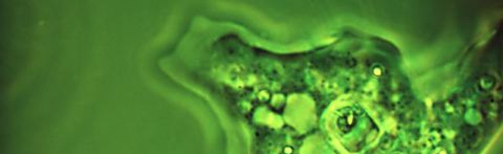
The cytoplasm (endoplasm is cytoplasm within the inner bit of the cell) is the gel-like substance within A. proteus that the organelles are suspended in. It is also the part of the cell that allows A. proteus to form its pseudopodia and preform its respective functions. The appearance of the cytoplasm in A. proteus is slightly granular – caused by tiny crystals in the cytoplasm. See the image below.

Note the granular appearance of the cytoplasm in A. proteus.
A. proteus (and amoebae in general) does not have a nervous system, nor sensory organelles. Thus, they solely rely on their cytoplasm for sensitivity.They repond to various stimuli such as: thigmotaxis, and phototaxis. The diagram below illustrates A. proteus reacting to various stimuli.
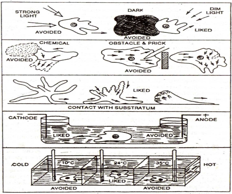
A. proteus reacting to various stimuli
Respiration:
A. proteus is an organism that requires oxygen, like other aerobic eukaryotes. It obtains oxygen through cellular respiration – basically the intake of oxygen and output of carbon dioxide. These gases simply enter and exit A. proteus VIA diffusion, through its semi-permeable membrane.
Plasma membrane:
This is a very thin membrane, with good regenerative abilities and elasticity. It contains the inner part of the cell (organelles, cytoplasm, etc.) and is semi permeable. It allows the movement of materials in and out of the cell (i.e. water, oxygen, waste, etc.), making it an important component of the cell. A feature that the plasma membrane of A. proteus has is the fact that it has many microvilli attached to it (can be seen under an electron microscope), which prevent the amoeba from sticking to the surface of the water.
Encystment:
When A. proteus “detects” unpleasant conditions – such as a nutrient defficient environment, it withdraws its pseudopodia and releases a protective covering over its plasma membrane (made of a chitin-like substance) called a cyst. This process often leads to one of the ways A. proteus reproduces – multi fission. This is one of the ways that A. proteus protects itself and ensures that it can reproduce under un-ideal conditions.
